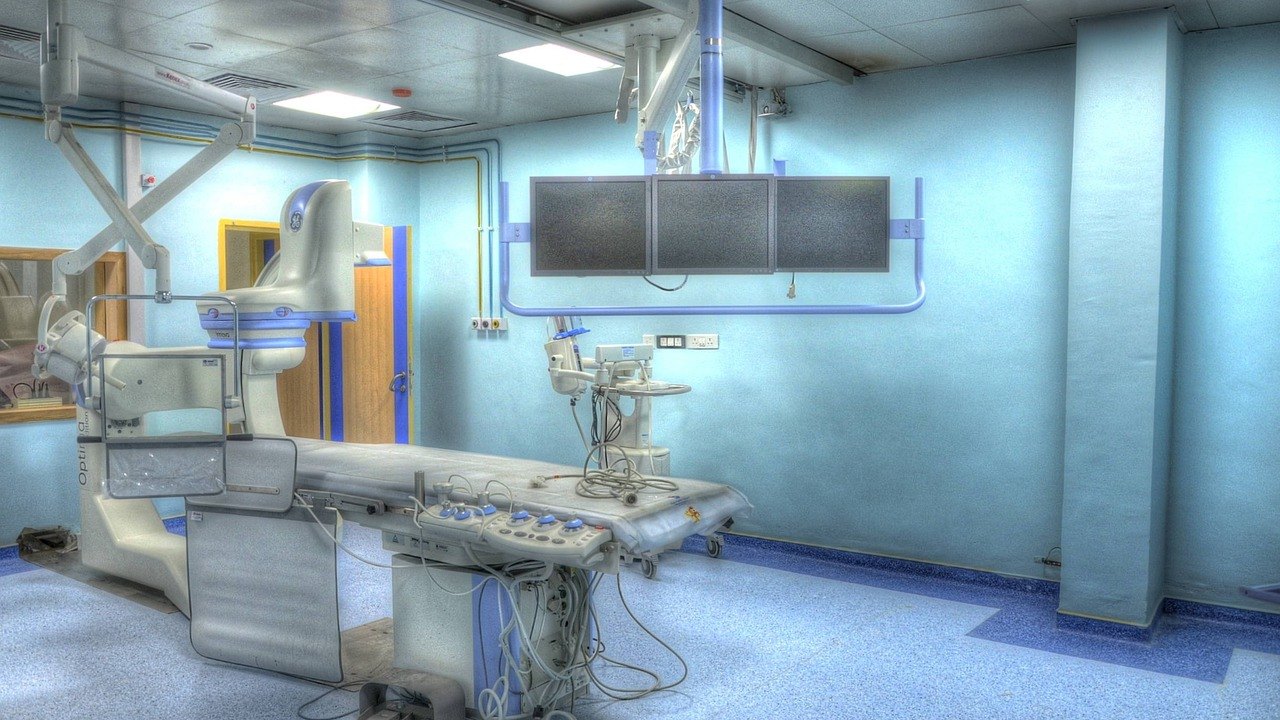
Atherosclerosis Catheter FLIm PS-OCT: First-in-Human Study
Researchers at the UC Davis National Center for Interventional Biophotonic Technologies (NCIBT) have finished the first-in-human test of a flexible coronary catheter that combines two optical imaging methods to look at atherosclerotic plaque. The device was evaluated in 10 patients under an FDA investigational device approval, and imaging of the last patient was completed in August. This is an early feasibility step: the source reports no data on effectiveness or on whether the device can predict a heart attack.
What happened
The catheter was developed in an academic lab rather than by a device company, which the team points out is unusual for a first-in-human study. It combines two technologies:
- Fluorescence lifetime imaging (FLIm), which measures the faint natural fluorescence of the artery wall. This reflects its biochemical composition.
- Polarization-sensitive optical coherence tomography (PS-OCT), which measures light scattered back from the same tissue to map its microstructure.
Both signals are acquired in one pullback. The catheter travels through the coronary vessels, rotates, and is withdrawn while it records live images. According to the report, it images an 80-millimeter length of coronary artery in about two seconds. The output is color images that show both structure and composition.
Before this study, the device was tested in animal models and in samples of human coronary arteries. The team's next step is to scale up to more patients. Jeffrey Southard, an interventional cardiologist, led the patient testing. Postdoctoral researcher Xiangnan Zhou operated the system in the cath lab.
Why it matters
Current cath lab imaging, such as intravascular ultrasound (IVUS) and conventional optical coherence tomography (OCT), shows the structure and microstructure of the vessel. According to the source, it does not show biochemical composition, which plays an important role in plaque instability and rupture.
The investigators say the new catheter can show plaque even before it causes stenosis. They hope it will eventually help clinicians judge plaque composition and pathobiology. That information could then inform how stents are placed and which drugs are used. Southard notes that further research is needed to learn the best ways to treat different plaque types.
These are stated goals, not demonstrated results. The report does not say whether the biochemical signal actually predicts rupture or erosion, or whether it improves outcomes.
What to know for the USMLE: atherosclerosis, vulnerable plaque, and intravascular imaging
The news is a good hook for several high-yield concepts. The following is standard textbook knowledge, not findings from the study.
- Pathogenesis: Atherosclerosis starts with endothelial injury or dysfunction (hypertension, smoking, hyperlipidemia, diabetes, shear stress). LDL enters the intima and is oxidized. Macrophages engulf it and become foam cells, forming the fatty streak. Smooth muscle cells migrate and lay down collagen, forming a fibrous cap over a lipid-rich necrotic core.
- Vulnerable plaque: The classic rupture-prone lesion is a thin-cap fibroatheroma. It has a large lipid core, a thin fibrous cap, and many inflammatory cells. Macrophages release matrix metalloproteinases, which degrade the collagen in the cap. This is why "biochemical composition" and inflammation matter, and why structure alone is an incomplete picture.
- From plaque to ACS: Plaque rupture exposes thrombogenic core material, which triggers platelet adhesion, aggregation, and thrombus formation. A partially occlusive thrombus typically produces unstable angina or NSTEMI. A fully occlusive thrombus produces STEMI. Plaque erosion (endothelial loss without cap rupture) is another substrate for thrombosis.
- Stenosis does not equal risk: Many MIs arise from plaques that were not severely stenotic beforehand. Outward (positive) remodeling, described as the Glagov phenomenon, can preserve the lumen while plaque grows. Stable angina is typically linked to fixed, flow-limiting stenosis (often cited around 70% or more), which causes demand ischemia on exertion.
- Why angiography has limits: Coronary angiography is a lumenogram. It shows the lumen silhouette, not the wall. IVUS and OCT image the wall itself. IVUS uses sound waves and penetrates deeper. OCT uses near-infrared light and has higher resolution but less penetration.
- Polarization-sensitive imaging: PS-OCT can detect birefringent tissue such as collagen. This is a basic physical principle and fits the cap-thickness and collagen story above. Fluorescence methods exploit tissue autofluorescence.
- Stent considerations: Stent placement is followed by dual antiplatelet therapy (aspirin plus a P2Y12 inhibitor such as clopidogrel). Complications to know are stent thrombosis (early) and in-stent restenosis (neointimal hyperplasia, later). Underexpansion and malapposition are recognized reasons to use intravascular imaging during stenting.
- Risk-factor and drug links: Statins (HMG-CoA reductase inhibitors) reduce events partly by lowering LDL and stabilizing plaques. The LAD is the most commonly occluded coronary artery in MI.

Limitations
- Very small, early study. Ten patients under an investigational device approval is a first-in-human feasibility step. The source gives no numbers on image quality, safety events, or diagnostic accuracy.
- No outcome data. The source states that the device's value for predicting plaque rupture and guiding treatment remains unestablished. No one has shown that biochemical imaging changes patient outcomes.
- Statements are expectations. Quotes such as the catheter providing information that "may indicate" inflammation and rupture risk describe a hope that needs validation in larger studies.
- Not available for routine care. The catheter is an investigational device. Standard practice continues to rely on existing tools such as angiography, IVUS, and OCT.
- Source type. The information comes from a news report based on the team's own statements, not a peer-reviewed results paper that we could examine.
FAQ
What is a dual-imaging catheter for atherosclerosis?
It is a catheter placed inside a coronary artery that collects two kinds of optical information in one pullback. FLIm gives biochemical information about the artery wall. PS-OCT gives microstructural information.
Can this catheter predict a heart attack?
That has not been shown. The researchers hope it will identify plaque features linked to rupture or erosion. The source says its value for predicting rupture and guiding treatment remains unestablished.
How is this different from IVUS or regular OCT?
IVUS and conventional OCT show vessel structure and microstructure. According to the source, they do not show biochemical composition. The new device aims to add that layer, and to do so together with microstructure in a single pass.
Key takeaways
- UC Davis completed the first-in-human test of a coronary catheter combining FLIm and PS-OCT in 10 patients. The device images 80 mm of artery in about 2 seconds.
- The goal is to capture plaque biochemistry plus microstructure. This addresses a gap in current imaging, but no efficacy or prediction data have been reported, and the next step is a larger study.
- For the USMLE, focus on the underlying concepts: thin-cap fibroatheroma, plaque rupture leading to ACS, the limits of angiographic stenosis, and the role of IVUS and OCT in the cath lab.
Prepared by the project team from the source material. Not medical advice.