Augmented Reality Liver Surgery: USMLE Couinaud Review
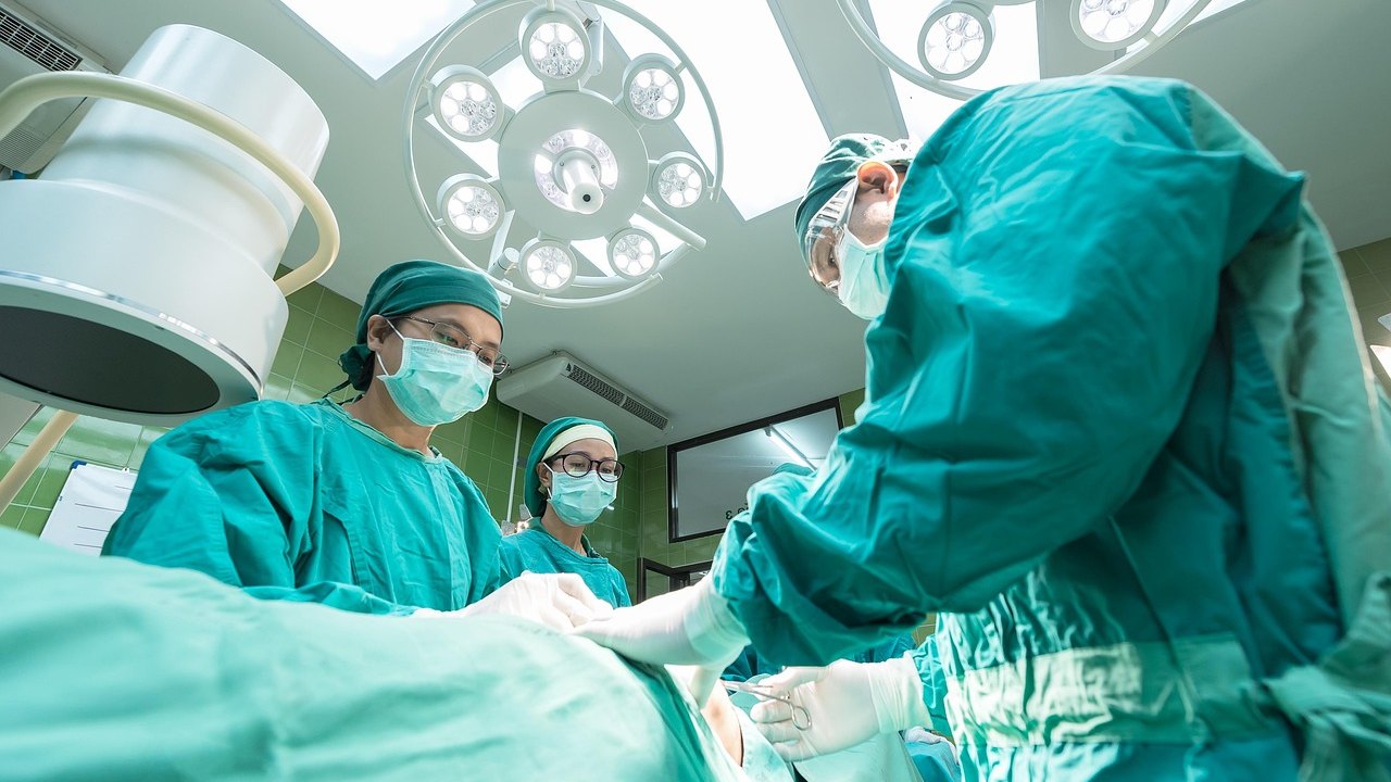
Researchers in Sweden have developed an augmented reality (AR) system that overlays a 3D model of a liver tumor and nearby structures onto the surgeon's laparoscopic camera view. The image updates as the camera moves. The system was used in surgery on a patient for the first time in June 2026, and the team reports it worked as planned. No data on patient outcomes have been described.
What happened
Tumors lying beneath the liver surface are hard to find during laparoscopic (keyhole) surgery. The surgeon sees only the organ's outer surface on a screen and cannot palpate it the way they would in open surgery. The new technology tries to solve this by showing where the tumor would appear "if the liver were semitransparent," as Torbjörn Lundh, a professor of mathematical sciences at Chalmers University of Technology and the University of Gothenburg, put it.
The method works like this, based on the source description:
- The surgeon places a sticker about one square centimeter (0.16 square inches) in size on the liver surface. It carries a QR code.
- The sticker serves as an artificial landmark on an otherwise smooth surface. Lundh compared the liver surface to a polished floor.
- The system combines the marker, the camera's position, and an X-ray image taken before surgery to calculate where the tumor should appear. It then overlays that information on the camera view.
- As long as the camera can see the marker, the system can display the tumor or other structures beneath the surface.
Niclas Kvarnström, a transplant surgeon and head of the Transplant Centre at Sahlgrenska University Hospital, described it as "a GPS inside the organ." He said the system was used as planned during the operation, the workflow worked well, and the patient went home the following day.
Further liver operations are planned for fall 2026. Work continues on clinical validation, product development, quality management systems, and the steps needed for regulatory approval. A larger international study is planned with the Intervention Centre at Oslo University Hospital and hospitals in Washington. The team also wants to extend the method to other laparoscopic procedures.
Why it matters
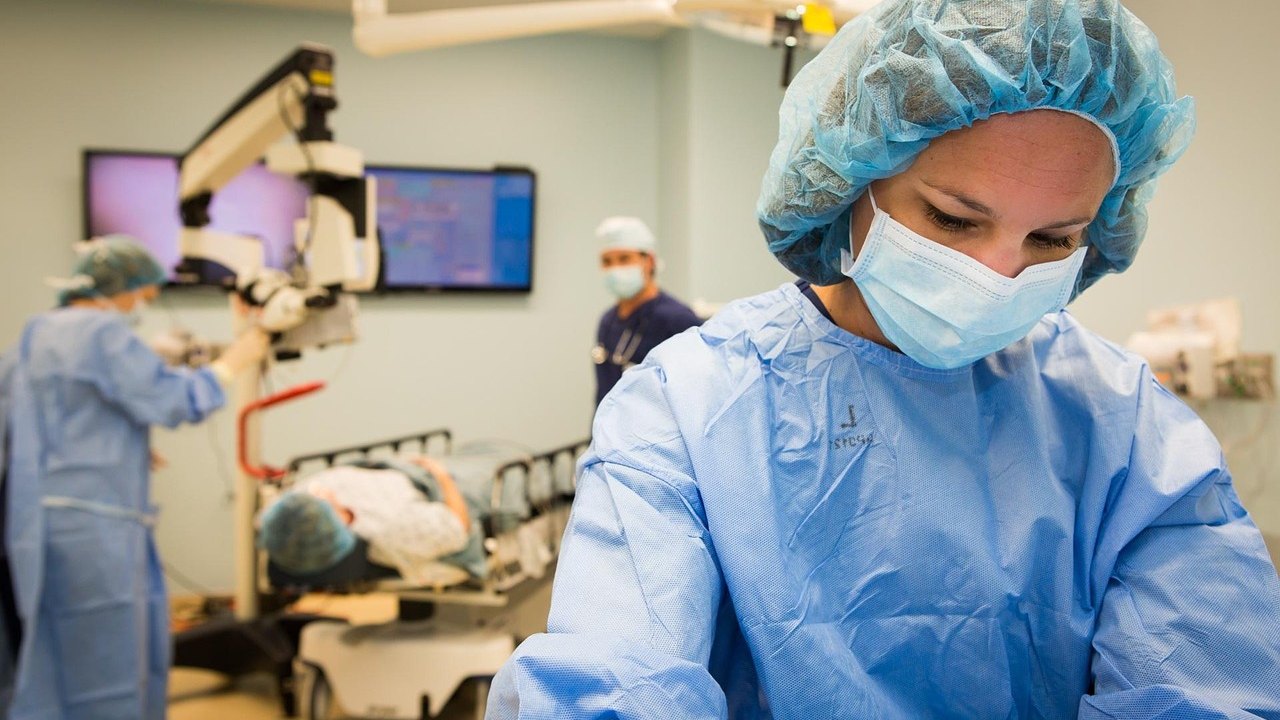
Laparoscopic surgery has well-known advantages, such as smaller incisions. Its main trade-off is the loss of direct touch and a limited, two-dimensional view. In liver surgery, that matters because a deep tumor cannot be seen or felt from the surface. Surgeons have to relate what they see to preoperative imaging in their heads, often with the help of intraoperative ultrasound.
An AR overlay aims to make that mental translation explicit and continuous. The idea is not new in principle. What the researchers describe is a specific mathematical solution that uses a small surface marker and updates with camera movement.
It is important to be clear about what this news does not show. The source describes one first-in-human use and a smooth workflow. It does not report whether the tumor was removed more accurately, whether margins improved, or whether complications decreased. For now, this is a feasibility milestone, not evidence of better outcomes.
What to know for the USMLE — high-yield liver anatomy and laparoscopy
The news is a good prompt to review the anatomy and physiology that a liver surgeon must navigate. Neither the news source nor the researchers made these points. They are standard textbook material.
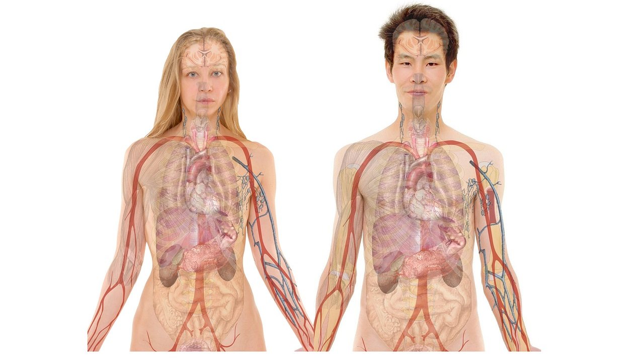
- Couinaud classification: The liver is divided into eight functionally independent segments (I–VIII). Each has its own portal triad (branches of the portal vein, hepatic artery, and bile duct) in the center. Hepatic veins run between segments.
- Cantlie line: It runs from the gallbladder fossa to the inferior vena cava and separates the functional right and left hemilivers. The middle hepatic vein runs along this plane.
- Segment I (caudate lobe): It drains directly into the IVC through short hepatic veins. It is often spared in Budd-Chiari syndrome (hepatic vein thrombosis), which causes caudate hypertrophy.
- Falciform ligament: It separates the left lateral section (segments II and III) from the left medial section (segment IV, the quadrate lobe).
- Portal triad and the Pringle maneuver: Clamping the hepatoduodenal ligament temporarily occludes the hepatic artery and portal vein to limit bleeding during resection. The triad passes anterior to the epiploic foramen (of Winslow).
- Liver tumors: In the Western world, metastases (classically from colorectal cancer) are the most common hepatic malignancy. The most common primary malignancy is hepatocellular carcinoma (HCC), linked to cirrhosis, hepatitis B and C, and elevated alpha-fetoprotein. Benign lesions include hemangioma and hepatic adenoma (associated with oral contraceptives).
- Why segments matter in resection: Surgeons often remove tumors along segmental planes to spare as much functional liver as possible. The liver can regenerate, but a sufficient future liver remnant is essential.
- Laparoscopy physiology: CO₂ pneumoperitoneum raises intra-abdominal pressure. This can reduce venous return, raise systemic vascular resistance, and cause hypercapnia and respiratory acidosis. Shoulder pain after laparoscopy is referred pain from diaphragmatic irritation through the phrenic nerve (C3–C5).
- Laparoscopy complications: Know trocar injury to vessels or bowel, CO₂ gas embolism, and subcutaneous emphysema.
Limitations
- One patient. The only clinical use reported is a single operation. One uneventful case shows the system can be used in the operating room, not that it works reliably.
- No outcome data. Nothing in the description addresses tumor localization accuracy, resection margins, operative time, complications, or recurrence. The statement that the patient went home the next day is an anecdote from one case, and it cannot be credited to the technology.
- Early stage. Clinical validation and regulatory approval are still in progress. The larger international study is only planned.
- Source type. The report is a news article built largely on statements from the developers. The references it cites are two 2020 master's theses, not a peer-reviewed clinical trial.
- Open technical questions. The source does not say how the system handles liver movement or deformation during surgery. The surface marker and camera position are used to calculate the tumor's location from imaging taken before the operation. Whether this remains accurate throughout a procedure is a question for future studies.
- Unspecified details. The source does not state the tumor type in the first patient or the exact imaging modality, referring only to an "X-ray image" taken before surgery.
FAQ
What is augmented reality in laparoscopic liver surgery?
It is a technique that overlays a computer-generated 3D model of the tumor and nearby anatomy onto the live camera image. In this system, a small QR-code sticker on the liver surface, the camera position, and preoperative imaging are used to calculate where the tumor lies beneath the surface.
Does this AR technology improve surgical outcomes?
That is not known yet. The source reports that the system was used as planned in one patient and that the workflow worked well. It gives no data on outcomes such as accuracy, margins, or complications. Larger studies are planned.
Is the technology approved for routine use?
No. According to the source, work continues on clinical validation, quality management systems, and the steps required for regulatory approval. More liver operations are planned for fall 2026.
Key takeaways
- A Swedish team has used an AR system for the first time in a liver operation (June 2026). It overlays a 3D tumor model on the laparoscopic view, using a roughly 1 cm² QR-code sticker on the liver surface.
- The evidence so far is one patient and a workflow report. There are no outcome data, and validation and regulatory approval are still pending.
- For the USMLE, use this as a prompt to review Couinaud segments, the Cantlie line, the portal triad and Pringle maneuver, common liver tumors, and the physiology and complications of laparoscopy.
Prepared by the project team from the source material. Not medical advice.