REDMOD AI Pancreatic Cancer Detection: USMLE Review
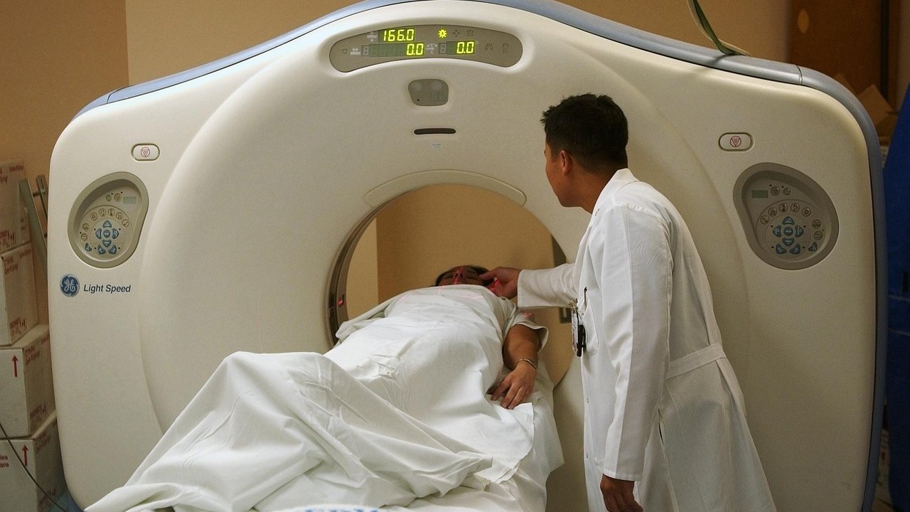
An artificial intelligence model called REDMOD detected subtle imaging signatures of pancreatic ductal adenocarcinoma (PDAC) on CT scans an average of 475 days before clinical diagnosis. These were scans that experienced radiologists had read as showing no cancer. The study, published in the journal Gut, reports that the AI had a sensitivity of 73%, compared with 39% for radiologists. It is a retrospective-style algorithm evaluation, not a ready-to-use screening test.
What happened
REDMOD stands for Radiomics-based Early Detection MODel. It analyzes very fine texture patterns in pancreatic tissue (radiomics) that are not readily visible on conventional CT. It also automatically outlines the pancreas and separates it from neighboring organs. That removes manual tracing by specialists, which can vary from person to person.
The researchers examined abdominal CT scans from 219 patients treated at multiple hospitals. At the time of imaging, radiologists had found no evidence of pancreatic cancer, but every one of these patients was later diagnosed with it. The timing of the scans before diagnosis was:
- 87 patients (40%): 3 to 12 months before diagnosis
- 76 patients (35%): 12 to 24 months before
- 56 patients (25%): more than 24 months before, up to about 3 years
In 64% of patients the tumor was in the head of the pancreas. The comparison group was 1243 people who stayed free of pancreatic cancer for up to three years afterward, matched by age, sex, and scan date. Mean age was 69 in the cancer group (range 34–88) and 64 in the comparison group (range 34–88).
The main results:
- Lead time: REDMOD identified cancer-associated signatures an average of 475 days before diagnosis.
- Sensitivity: 73% for the AI vs 39% for radiologists.
- Scans more than 2 years before diagnosis: 68% for the AI vs 23% for radiologists.
- Cancer-free scans: In an independent group of 539 patients from several hospitals, just over 81% were correctly classified as cancer free. On the public NIH-PCT dataset (80 patients), 87.5% were correctly identified as cancer free.
- Reproducibility: On scans from the same patients taken several months apart, results matched in 90–92% of cases.

Why it matters
PDAC has a low survival rate, largely because it is usually found late. Early disease causes few noticeable symptoms, and the tissue changes can be too subtle for standard imaging. The cancer can also progress quickly.
The authors cite modeling studies suggesting that raising the share of localized PDAC from 10% to 50% would more than double survival. They describe diagnosis timing as the most critical determinant of outcome. They also describe what REDMOD detects as imaging signatures of "stage 0" disease in a pancreas that looked normal. That is the researchers' interpretation, and it still needs prospective confirmation.
The team stresses that the next step is testing in high-risk groups, such as people with unexplained weight loss or recently diagnosed diabetes.
What to know for the USMLE — PDAC, imaging, and screening logic
Use this news as a hook to review pancreatic cancer and basic biostatistics:
- Epidemiology and pathology: PDAC is the most common pancreatic cancer. It arises from precursor lesions called PanIN. Molecular steps classically include KRAS activation (very common), then CDKN2A (p16), TP53, and SMAD4 inactivation.
- Risk factors: Smoking is the major modifiable factor. Others include chronic pancreatitis, obesity, older age, and new-onset diabetes in older adults. Hereditary syndromes include BRCA2, CDKN2A (familial atypical multiple mole melanoma), STK11 (Peutz-Jeghers), Lynch syndrome, and hereditary pancreatitis (PRSS1).
- Classic presentation: Weight loss, epigastric pain radiating to the back, and painless jaundice with a head tumor. A palpable, nontender gallbladder is Courvoisier sign. Trousseau syndrome (migratory thrombophlebitis) is a paraneoplastic hypercoagulable state. Steatorrhea can occur from duct obstruction.
- Head vs body/tail: Head tumors obstruct the common bile duct early and cause jaundice. Body and tail tumors often present later and with more advanced disease.
- Imaging: The first test in suspected PDAC is a pancreatic-protocol (multiphase) CT. Typical findings are a hypoattenuating mass and the double-duct sign (dilated bile and pancreatic ducts). EUS with fine-needle aspiration is used for tissue diagnosis, and MRI/MRCP is a complement. REDMOD works on routine CT, which is why its "invisible" signal is notable.
- Tumor marker: CA 19-9 is used to follow treatment response and recurrence. It is not a good screening test because it lacks sensitivity and specificity and can be elevated in biliary obstruction.
- Treatment concepts: Resectable head tumors are treated with the Whipple procedure (pancreaticoduodenectomy). Chemotherapy such as FOLFIRINOX is used in many settings. Few patients present with resectable disease, which is why early detection matters.
- Biostatistics:
- Sensitivity = TP / (TP + FN). It is the ability to catch true cases (73% vs 39% here).
- Specificity = TN / (TN + FP). The cancer-free classification rates (about 81% and 87.5%) reflect this idea.
- Predictive values depend on prevalence. PDAC is rare in the general population, so even a decent specificity can produce many false positives. This is why screening is usually proposed for high-risk groups.
- Lead-time bias: Earlier detection can make survival look longer without changing the time of death. Only outcome trials can show true benefit.

Limitations
- Selected, case-control-style design. The cancer group consisted of people who were all later diagnosed, compared with matched people who remained cancer free. This is not the same as screening an unselected population, where most people never develop PDAC.
- No prospective testing yet. The authors say prospective validation is required to confirm clinical usefulness, especially in people at increased risk.
- Limited diversity. The study population did not include a sufficiently diverse range of ethnic backgrounds, so performance in other populations is uncertain.
- Age difference between groups. The cancer group averaged 69 years and the comparison group 64, even though matching by age was reported.
- Specificity leaves room for false positives. Just over 81% and 87.5% correct cancer-free classifications in the two test sets mean a meaningful fraction of cancer-free scans were not recognized as such. What that means for follow-up testing and patient anxiety would need real-world study.
- The "stage 0" label is an interpretation. The findings show a statistical imaging signal that preceded diagnosis, and the source gives no confirmation of the underlying pathology on those earlier scans.
This article is educational and does not offer advice about testing or care for any individual.
FAQ
What is REDMOD?
It is an AI framework (Radiomics-based Early Detection MODel) that analyzes texture patterns in the pancreas on CT scans and automatically outlines the organ. In the study it flagged signs of pancreatic ductal adenocarcinoma before radiologists could see them.
Is REDMOD available for pancreatic cancer screening?
No. The researchers say prospective validation is needed, particularly in high-risk groups such as people with unexplained weight loss or recently diagnosed diabetes. The current results come from algorithm testing on existing scans.
Why is pancreatic cancer usually diagnosed late?
Early disease causes few symptoms, and the tissue changes can be too subtle for standard imaging. The cancer can also progress quickly. Many patients are therefore diagnosed at an advanced stage, when survival is low.
Key takeaways
- REDMOD detected cancer-associated CT signatures a mean of 475 days before diagnosis, with 73% sensitivity vs 39% for experienced radiologists. Correct cancer-free classification was just over 81% and 87.5% in two test sets.
- The evidence is retrospective and not yet tested prospectively, and the study population lacked ethnic diversity, so this is not a screening tool yet.
- For the USMLE, tie this to PDAC risk factors, presentation (painless jaundice, Courvoisier, Trousseau), CT as first-line imaging, CA 19-9 limits, and the sensitivity, specificity, predictive value, and lead-time bias concepts.
Prepared by the project team from the source material. Not medical advice.